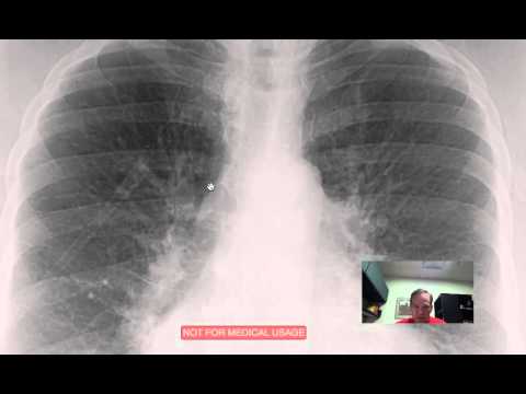
Pulmonary plethora is a term used to describe the appearances of increased pulmonary perfusion on chest radiographs. It is commonly used in pediatric radiology.
Q. What does asunder mean?
into parts torn
Table of Contents
- Q. What does asunder mean?
- Q. What is another word for plethora?
- Q. What is a Hampton’s hump?
- Q. What is pulmonary Oligemia?
- Q. What is pulmonary perfusion?
- Q. What is a VQ medical test?
- Q. What is the difference between perfusion and diffusion?
- Q. Can you eat before VQ scan?
- Q. How accurate is a VQ scan?
- Q. What can I expect from a VQ scan?
Q. What is another word for plethora?
Plethora Synonyms – WordHippo Thesaurus….What is another word for plethora?
| excess | overabundance |
|---|---|
| superfluity | surfeit |
| surplus | profusion |
| superabundance | glut |
| abundance | cornucopia |
Q. What is a Hampton’s hump?
Hampton’s hump is a radiological sign consisting of a peripheral, wedge-shaped opacification adjacent to the pleural surface, which represents pulmonary infarction distal to a pulmonary embolus. 1. Owing to good pulmonary perfusion from collateral blood vessels, this sign is rarely seen in clinical practice.
Q. What is pulmonary Oligemia?
Westermark sign is a chest x-ray finding of oligaemia (clarified area) distal to a large vessel that is occluded by a pulmonary embolus. The focal area of increased translucency due to oligaemia is caused by impaired vascularisation of the lung due to primary mechanical obstruction or reflex vasoconstriction.
Q. What is pulmonary perfusion?
Gas exchange occurs in the lungs between alveolar air and blood of the pulmonary capillaries. For effective gas exchange to occur, alveoli must be ventilated and perfused. Ventilation (V) refers to the flow of air into and out of the alveoli, while perfusion (Q) refers to the flow of blood to alveolar capillaries.
Q. What is a VQ medical test?
Also known as Lung or Pulmonary Ventilation (V) and Perfusion (Q) Scans. Leer en español. A lung VQ scan is an imaging test that uses a ventilation (V) scan to measure air flow in your lungs and a perfusion (Q) scan to see where blood flows in your lungs.
Q. What is the difference between perfusion and diffusion?
The main difference between perfusion and diffusion is that perfusion is the delivery of blood to the pulmonary capillaries, whereas diffusion is the movement of gases from the alveoli to plasma and red blood cells. Here, the gas exchange of animals occurs through the respiratory membrane of the alveoli of the lungs.
Q. Can you eat before VQ scan?
Generally, there is no special dietary preparation, such as fasting, before the scan. You may also be asked to have an X-ray of your chest done 24 to 48 hours before your test.
Q. How accurate is a VQ scan?
With such a high specificity, if the test is positive, the diagnosis can be ruled in or confirmed. Several papers have reported statistically significant greater accuracy for PE detection for CT with sensitivities and specificities for CT of 83% to 94% and 94% to 96%, respectively vs. 65% and 94% for V/Q scintigraphy.
Q. What can I expect from a VQ scan?
A VQ scan is carried out in two parts. In the first part, radioactive material is breathed in and pictures or images are taken to look at the airflow in the lungs. In the second part, a different radioactive material is injected into a vein in the arm, and more images taken to see the blood flow in the lungs.