Structures that are dense (such as bone) will block most of the x-ray particles, and will appear white. Metal and contrast media (special dye used to highlight areas of the body) will also appear white. Structures containing air will be black, and muscle, fat, and fluid will appear as shades of gray.
Q. How can the amount of scatter radiation reaching the IR be controlled?
The radiographer must act to minimize the amount of scatter radiation reaching the IR. Beam-restricting devices and radiographic grids are tools the radiographer can use to limit the amount of scatter radiation that affects the radiographic image and exposure to the patient or personnel.
Table of Contents
- Q. How can the amount of scatter radiation reaching the IR be controlled?
- Q. How is Sid related to exposure rate and image density?
- Q. What happens when you increase mAs?
- Q. What is fluoroscopic imaging?
- Q. What causes foreshortening?
- Q. How do you fix a bitewing overlap?
- Q. What is the result of incorrect horizontal angulation?
- Q. What is bitewing technique?
- Q. Which contact should you open for a premolar bitewing?
- Q. What landmarks should be seen on a bitewing radiograph?
Q. How is Sid related to exposure rate and image density?
As SID increases, exposure rate decreases and radiographic density decreases. A radiograph of the lumbar spine requires an exposure of 70 kVp and 320 mAs.
Q. What happens when you increase mAs?
An increase in current (mA) results in a higher production of electrons that are inside the x-ray tube which will, therefore, increase the quantity of radiation; more radiation will cause more photons reaching the detector and hence apparent structural density will decrease, yet the signal intensity will increase.
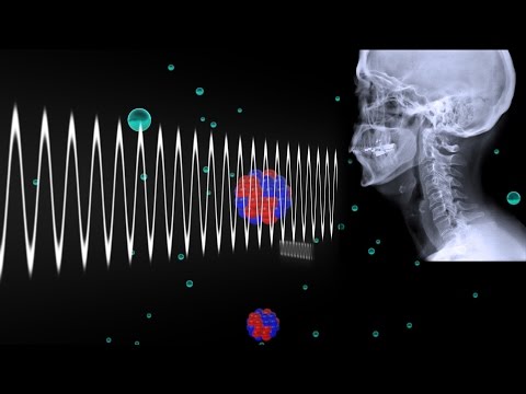
Q. What is fluoroscopic imaging?
Fluoroscopy is a type of medical imaging that shows a continuous X-ray image on a monitor, much like an X-ray movie. During a fluoroscopy procedure, an X-ray beam is passed through the body.
Q. What causes foreshortening?
Foreshortening is the result of overangulation of the x-ray beam. When foreshortening occurs when using the paralleling technique, the angulation of the x-ray beam is greater than the long axis plane of the teeth. This error can also occur if the receptor is not placed parallel to the long axis of the teeth.
Q. How do you fix a bitewing overlap?
Horizontal overlap is a result of the X-ray beam not passing through the open interproximal area at right angles to a properly positioned detector. Correcting this error on bitewings can usually be achieved by inclining the tubehead in a more mesial or distal direction.
Q. What is the result of incorrect horizontal angulation?
Incorrect horizontal angulation results in overlapped (unopened) contact areas. A film with overlapped contact areas cannot be used to examine the interproximal areas of the teeth.
Q. What is bitewing technique?
Bitewing Technique The bitewing radiographic image is used to examine the interproximal surfaces of the teeth and is particularly useful for the detection of dental caries and alveolar bone levels. The receptor is placed into the mouth parallel to the crowns of the maxillary and mandibular posterior teeth.
Q. Which contact should you open for a premolar bitewing?
The central x-ray beam should be parallel to the interproximal spaces. This will eliminate the chances of overlap and ensure open contacts. For the premolar bitewing, it is expected that the distal of the canines are present.
Q. What landmarks should be seen on a bitewing radiograph?
Bitewing radiographs record, on a single image, the crowns and coronal 1/3 of the interproximal bone of both arches. Bitewings are useful for detecting interproximal carious lesions, bone height, pulp chamber size and shape, pulp stones, and overhangs on interproximal restorations.